December 04, 2024

The most complicated object in the known Universe is bound to inspire some heated debate, but neuroscientists are now arguing over a basic aspect of the brain we thought we'd figured out.
A controversial study, led by Jacqueline Griswold from Johns Hopkins University in the US, has some scientists arguing for a fundamental change in how we view neurons – the building blocks of the brain and nervous system.
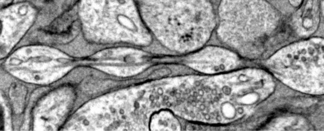
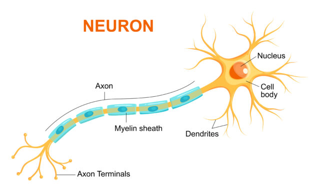
Despite what most diagrams show, axons – the main arms of neurons – are not a smooth cylinder, they say, but more like a string of pearls. The size and spacing of those nanoscopic bumps are dynamic, possibly controlling how quickly messages are sent in the brain.
"Understanding the structure of axons is important for understanding brain cell signaling," explains molecular neuroscientist Shigeki Watanabe, who is head of the lab at Johns Hopkins.
"Axons are the cables that connect our brain tissue, enabling learning, memory, and other functions. These findings challenge a century of understanding about axon structure."

It's a tiny detail that could have big repercussions, but other neuroscientists aren't buying it.
"I think it's true that [the axon is] not a perfect tube, but it's not also just this kind of accordion that they show," neuroscientist Christophe Leterrier from Aix-Marseille University told Sofia Quaglia at Science.
Previous studies have found that when brain cells are damaged or dying, the tails can start to 'bubble', creating a bead-like pattern. 'Axonal beading' is especially common in the brains of those with Alzheimer's or Parkinson's disease.

But Watanabe and his colleagues say the pearls they have found in the brains of mice are on a nanoscale, not a microscale like previous cases of axonal beading.
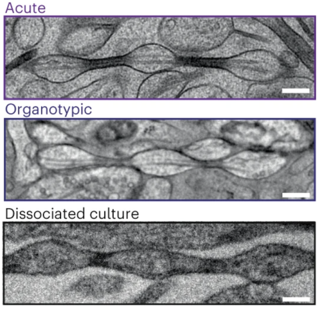
Analyzing the brain slices of mice at various ages, the team has zoomed in on individual axons without a protective sheath.
No matter how the brain tissue slices were cultured, axons did not look smooth but were riddled with 'nanopearls' of various sizes.
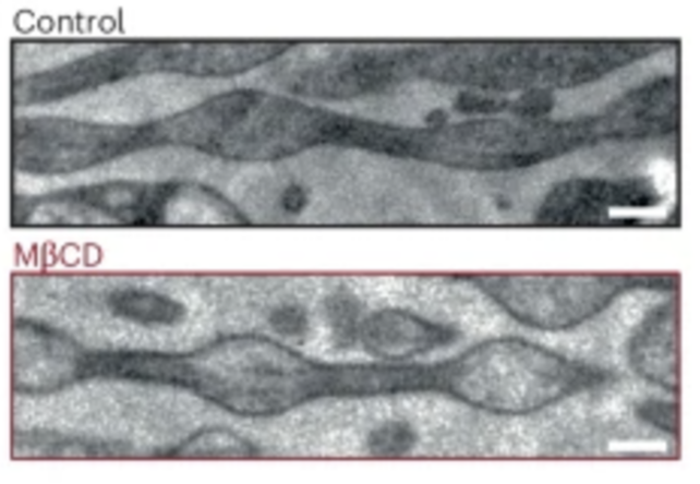
What's more, the size of these pearls could be manipulated with predictive outcomes. Removing cholesterol from the axon, for instance, resulted in less pearling and a reduced ability to send electrical messages.

But some critics think the nanopearls seen in mouse neurons are responses to the stress of tissue culturing.
Previous studies have found that when stretched, axons can form macro beads that are sort of like "stress balls for the brain". These clumps might form to stop the spread of damaging wave signals through a neuron's tail, and they tend to somewhat resolve after about 15 minutes.
If culture techniques stress a mammalian neuron, then some experts believe it is possible nanopearls will form. But if that occurs, this would be a response to stress, not a healthy state.
Lead author Griswold told Science that is why her team also imaged live cells that were not frozen or chemically affixed. These, too, showed the nanopearl pattern.

In Griswold's experiments, the chemical that is usually used for imaging neurons caused the nanopearls to disappear, possibly explaining why they haven't been seen before.
That said, some scientists have seen similar nanopearling in the axons of comb jellies, and in the past, Watanabe has regularly noticed the effect in roundworm axons, too.
More evidence is the only way to end the debate once and for all. Watanabe and his colleagues at Johns Hopkins are now studying the neurons from human brains to see if they can find more nanopearls in a unit a hundred times smaller than the width of a hair.
The study was published in Nature Neuroscience.